October 5, 2026
EGFR Mutation Genotyping and SHOX2 / RASSF1A Lung Cancer Methylation Testing: A Two-Assay Molecular Workflow on Real-Time PCR
Lung cancer in never-smoking populations has become one of the more closely studied puzzles in thoracic oncology, and one line of evidence points to a disease that is molecularly distinct from the smoking-associated form. In 2020, a study published in Cell set out to characterise that disease directly.
The researchers behind “Proteogenomics of Non-smoking Lung Cancer in East Asia Delineates Molecular Signatures of Pathogenesis and Progression” conducted a deep proteogenomic analysis of a prospectively collected cohort of lung adenocarcinoma patients in Taiwan, representing early-stage, predominantly female, non-smoking disease. Their integrated approach combined genomic, proteomic and phosphoproteomic profiling of tumour tissue to define the molecular attributes of this population and to trace the hallmarks of tumour progression [4].
The central finding concerned driver mutations. EGFR mutations were the predominant driver alteration in this never-smoker cohort — the study's own summary describes the disease as being characterised by “predominant EGFR mutations,” in contrast to the far smaller share of EGFR-driven tumours found in smoking-associated lung cancer. Mutational signature analysis further revealed age- and gender-related mutagenesis mechanisms: a high prevalence of an APOBEC mutational signature in younger females, and an over-representation of environmental-carcinogen-like mutational signatures in older females. A proteomics-informed classification distinguished the clinical characteristics of early-stage patients with EGFR mutations, and integrated protein network analysis nominated candidate biomarkers for patient stratification and therapeutic intervention [4].
The study’s significance lies less in any single figure than in what it establishes about the disease itself: in this demographic, lung cancer is not simply a less common variant of the smoker’s disease but a biologically distinct entity with its own driver profile, its own mutagenic history and its own candidate biomarkers. That has a direct practical consequence. If EGFR drives a large share of these tumours, then identifying EGFR mutation status is not an optional refinement of a diagnosis — it is the step that determines whether a targeted therapy is available to the patient at all [6,8]. This technical article describes two CE-marked, real-time PCR assays — an EGFR mutation detection kit covering exons 18–21, and a dual-gene lung cancer methylation panel targeting SHOX2 and RASSF1A — and explains where each belongs in a modern thoracic-oncology laboratory workflow.
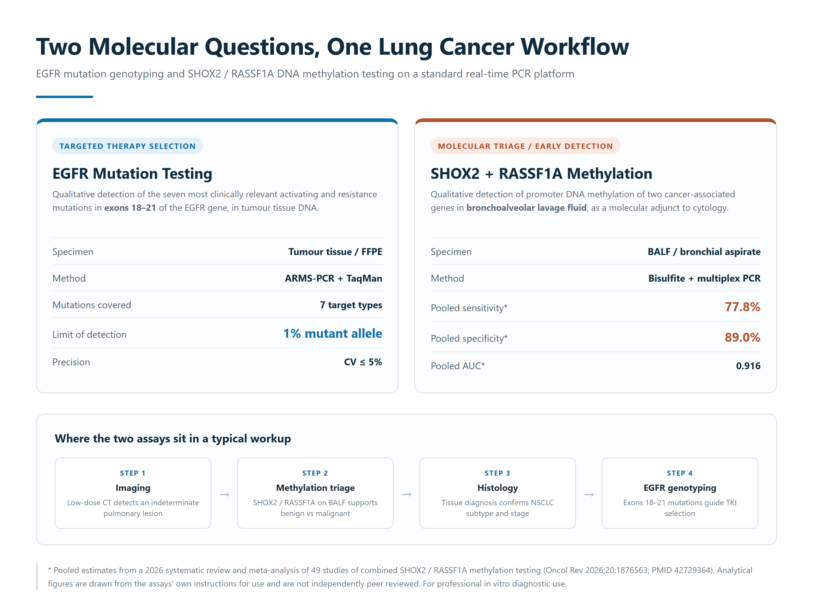
Key facts at a glance
- EGFR assay analytes: seven clinically relevant EGFR mutation types across exons 18, 19, 20 and 21 (G719X, DEL19, S768I, T790M, INS20, L858R, L861Q), reported qualitatively in tumour tissue DNA
- Methylation assay analytes: promoter DNA methylation of SHOX2 and RASSF1A, reported qualitatively in bronchoalveolar lavage fluid (BALF)
- Methylation method: bisulfite conversion followed by multiplex real-time PCR with hydrolysis (TaqMan) probes in a single closed tube
- EGFR method: ARMS-PCR combined with TaqMan hydrolysis probes, discriminating mutant from wild-type sequence at the primer level
- EGFR limit of detection: 1% mutant allele
- Methylation limit of detection: ≤ 250 copies per test
- Precision (both assays): Ct coefficient of variation ≤ 5%
- Methylation turnaround: approximately 6 hours from specimen to reported result
- Instrumentation: widely used real-time PCR platforms with FAM, VIC/HEX and CY5 channels
- Regulatory status: both kits are CE-marked in vitro diagnostics under Directive 98/79/EC; the methylation panel is also registered with China's National Medical Products Administration
The clinical problem: a shifting epidemiology and a detection gap
Lung cancer remains the world's most frequently diagnosed cancer and its leading cause of cancer death. GLOBOCAN 2022 estimates 2.48 million new cases and 1.80 million deaths worldwide, accounting for 12.4% of all cancer diagnoses and 18.7% of all cancer deaths [1]. The prognosis gap is a detection gap: early-stage disease is highly curable, while advanced-stage disease is not.
What has changed is who is being diagnosed. A 16-year hospital-based analysis of more than 24,000 surgically managed lung cancer patients, published in Lung Cancer in 2025, found the proportion of women in the surgical population rising from 32% in 2005 to 48% in 2021, with more than 70% of those women never having smoked, compared with fewer than 10% of men. The number of female patients under 40 doubled across the study period, and adenocarcinoma rose from roughly 60% to more than 80% of female cases [2].
A companion population-based cohort using regional electronic health records from Eastern China — covering more than 1.2 million people across Shanghai, Jiangsu and Zhejiang — found that lung cancer incidence among never-smoking women doubled over 13 years, from 9.2 to 18.6 per 100,000. In several age bands, including the under-40 and under-50 groups, female incidence exceeded that of men for the first time, with the steepest growth in women aged 35–50 [3]. These are not isolated case reports. They describe a demographic shift with direct consequences for how laboratories test for lung cancer.
Why EGFR matters: the targetable driver
EGFR (epidermal growth factor receptor) is a receptor tyrosine kinase whose activating mutations drive a substantial proportion of non-small cell lung cancer (NSCLC). The mutational landscape of never-smoker lung cancer in East Asia is distinct: a deep proteogenomic study of early-stage, predominantly female, non-smoking lung adenocarcinoma characterised a high prevalence of EGFR mutations alongside an APOBEC-associated mutational signature enriched in younger women, and environmental-carcinogen-like signatures in older women [4]. A global systematic review of EGFR mutation prevalence confirmed that mutation frequency varies sharply by population, making molecular testing — rather than clinical assumption — the only reliable way to identify who will respond to targeted therapy [5].
Detecting EGFR mutations is not academic. The IPASS trial established that EGFR mutation status determines whether a tyrosine kinase inhibitor (TKI) outperforms chemotherapy [6]; EURTAC extended the principle to erlotinib in European patients [7]; and FLAURA showed that first-line osimertinib achieved a median progression-free survival of 18.9 months versus 10.2 months for standard EGFR-TKIs (hazard ratio 0.46, 95% CI 0.37–0.57) [8], with a final overall survival analysis reporting a median of 38.6 versus 31.8 months [9]. Guidelines from the College of American Pathologists, IASLC and AMP, and from ESMO, all require EGFR testing at diagnosis in advanced non-squamous NSCLC [10,11]. A molecular assay is therefore the gateway to the entire class of EGFR-directed therapy.
Why DNA methylation matters: an early, complementary signal
DNA methylation of tumour-suppressor and homeobox gene promoters is an early and near-universal event in lung carcinogenesis. Because it is detectable before clinical symptoms and is mechanistically tied to gene silencing, methylated DNA functions as a methylation biomarker that can complement both imaging and cytology. Two genes are particularly well characterised:
- SHOX2 (short stature homeobox 2) is a homeobox transcription factor whose promoter is aberrantly methylated in several solid tumours. SHOX2 methylation distinguishes benign from malignant lung lesions and has been reported as both an early-detection and an independent prognostic marker in NSCLC.
- RASSF1A (Ras association domain family 1, isoform A) is a tumour suppressor silenced in tumours by promoter hypermethylation, loss of heterozygosity and chromosomal deletion. It contributes the highest specificity component of the pair.
The combined evidence base for this pair has now matured into a formal meta-analysis. A 2026 systematic review of 49 studies evaluated SHOX2 and RASSF1A methylation for lung cancer detection and reported a pooled sensitivity of 77.8% (95% CI 72.3–82.5%) and a pooled specificity of 89.0% (95% CI 86.6–91.1%), with an HSROC area under the curve of 0.916. SHOX2 alone yielded 69.4% sensitivity and 91.7% specificity; RASSF1A alone yielded lower sensitivity (45.7%) but the highest specificity (93.8%). The combined panel significantly improved sensitivity over either marker alone while retaining specificity comparable to SHOX2 [12]. The same pair has independently been meta-analysed in malignant pleural effusion, where adding methylation testing to cytology improved diagnostic accuracy for distinguishing malignant from benign effusions [13].
How the two assays work in practice
The EGFR mutation detection kit uses ARMS-PCR in combination with TaqMan hydrolysis probes. Allele-specific primers amplify only when perfectly matched to the target mutation sequence, allowing the assay to discriminate a low-abundance mutant allele from a large excess of wild-type DNA. Seven mutation types are covered in a single run: exon 19 deletions (DEL19), exon 21 L858R and L861Q, exon 18 G719X, exon 20 S768I, exon 20 insertions (INS20), and the acquired-resistance mutation T790M. Dual quality control — an internal standard and an external standard reaction — verifies DNA quality and PCR integrity in every run. The assay accepts fresh, frozen or formalin-fixed paraffin-embedded (FFPE) tumour tissue that has been confirmed to contain tumour cells, and is supplied at 24 tests per kit.
The methylation panel begins with bisulfite conversion, which chemically converts unmethylated cytosine to uracil while leaving methylated cytosine unchanged, so that the two states can be distinguished by sequence. Converted DNA is then amplified in a single closed tube using three fluorescent channels: FAM for methylated RASSF1A, VIC/HEX for methylated SHOX2, and CY5 for an internal control. Multiplexing both markers in one reaction conserves scarce BALF specimens and reduces hands-on time. The reported limit of detection is ≤ 250 copies per test, with ≤ 5% Ct coefficient of variation, and the closed-tube format limits the contamination risk that open post-PCR workflows carry.
Analytical performance
- EGFR assay — mutations detected: DEL19, L858R, L861Q, G719X, S768I, INS20, T790M, across exons 18–21 in a single run
- EGFR assay — limit of detection: 1% mutant allele frequency
- EGFR assay — precision: Ct coefficient of variation ≤ 5%
- EGFR assay — specimen types: fresh, frozen or FFPE tumour tissue with confirmed tumour cells
- Methylation panel — limit of detection: ≤ 250 copies per test
- Methylation panel — precision: Ct coefficient of variation ≤ 5%
- Methylation panel — specimen type: bronchoalveolar lavage fluid (BALF), bronchial aspirate or brush eluate
- Methylation panel — turnaround time: approximately 6 hours from specimen to reported result
All analytical figures above are taken from each product's own instructions for use and are the manufacturer's internal documentation. They are not independently peer reviewed and are presented as the developer's own performance specifications.
Clinical performance
| Assay | Specimen | Performance | Source |
|---|---|---|---|
| SHOX2 + RASSF1A methylation | Tissue and liquid biopsy (mixed) | Pooled sensitivity 77.8%; specificity 89.0%; AUC 0.916 | 49-study meta-analysis, 2026 [12] |
| SHOX2 methylation alone | Tissue and liquid biopsy (mixed) | Sensitivity 69.4%; specificity 91.7% | 49-study meta-analysis, 2026 [12] |
| RASSF1A methylation alone | Tissue and liquid biopsy (mixed) | Sensitivity 45.7%; specificity 93.8% | 49-study meta-analysis, 2026 [12] |
| SHOX2 + RASSF1A methylation | Pleural effusion | Improved accuracy over cytology alone for malignant vs benign effusion | Systematic review and meta-analysis, 2025 [13] |
| Methylation panel, as supplied | BALF | 75–81% sensitivity; 90–97% specificity | Developer's own clinical dossier, not independently peer reviewed |
| Methylation panel, as supplied | FFPE tissue | 89.8% sensitivity; 90.4% specificity | Developer's own clinical dossier, not independently peer reviewed |
Peer-reviewed evidence retrievable on PubMed
Every study below is retrievable on PubMed under the identifier given, and full citations with DOIs appear in the reference list.
Epidemiology of lung cancer in never smokers
- A 16-year hospital-based analysis of more than 24,000 surgically treated patients documented rising female representation, a doubling of patients under 40, and adenocarcinoma exceeding 80% of female cases. (PMID 40986991) [2]
- A population-based cohort of more than 1.2 million people in Eastern China found lung cancer incidence among never-smoking women doubling from 9.2 to 18.6 per 100,000, exceeding male incidence in several younger age bands. (PMID 40930563) [3]
- Proteogenomic profiling of early-stage, non-smoking lung adenocarcinoma in East Asia delineated APOBEC-associated and environmental-carcinogen mutational signatures and a high prevalence of EGFR mutations in younger women. (PMID 32649875) [4]
- A controlled multi-omics study of airborne pollutant exposure demonstrated a cross-organ pathway linking pulmonary inflammation to gastrointestinal and systemic inflammatory changes after short-term cooking-oil-fume inhalation. (PMID 39145585) [14]
EGFR as a therapeutic target
- IPASS established that EGFR mutation status determines whether a TKI outperforms chemotherapy: among 261 EGFR mutation-positive patients, progression-free survival was significantly longer with gefitinib than with carboplatin plus paclitaxel (hazard ratio 0.48, 95% CI 0.36–0.64, P<0.001), while mutation-negative patients did not show that benefit. (PMID 19692680) [6]
- EURTAC confirmed the same principle for erlotinib as first-line therapy in European patients with advanced EGFR mutation-positive NSCLC. (PMID 22285168) [7]
- FLAURA reported a median progression-free survival of 18.9 months with first-line osimertinib versus 10.2 months with standard EGFR-TKIs (hazard ratio 0.46, 95% CI 0.37–0.57). (PMID 29151359) [8]
- The final FLAURA overall survival analysis reported a median overall survival of 38.6 months with osimertinib versus 31.8 months with comparator EGFR-TKIs. (PMID 31751012) [9]
- A global systematic review and meta-analysis quantified the prevalence of EGFR mutations across NSCLC populations and subgroups, confirming substantial geographic variation. (PMID 27738317) [5]
DNA methylation as a lung cancer biomarker
- A 2026 systematic review and meta-analysis of 49 studies found that the combined SHOX2/RASSF1A methylation panel achieved a pooled sensitivity of 77.8% and specificity of 89.0% (AUC 0.916), outperforming either marker alone. (PMID 42729364) [12]
- A separate systematic review and meta-analysis evaluated SHOX2 and RASSF1A methylation in malignant pleural effusion, supporting methylation testing as an adjunct for effusion triage. (PMID 40614167) [13]
- A study comparing supernatants and matched cell pellets established the optimal specimen fraction for SHOX2/RASSF1A methylation testing in pleural effusion. (PMID 38072300) [15]
- A cell-free DNA methylation strategy using the SHOX2/RASSF1A panel demonstrated feasibility for distinguishing malignant from benign pleural effusion. (PMID 36691467) [16]
- A systematic review of methylated circulating tumour DNA in blood concluded that methylated ctDNA shows promise as a rule-in tool for lung cancer diagnosis, while noting its limitations as a rule-out test. (PMID 37568774) [17]
Evidence map: what the current documentation contains, and what a laboratory should request
Read this as an evidence map, not a performance claim. It sets out, for each assay, which evidence elements are present in the documentation supplied with the product and which a laboratory should request before committing to a tender or a validation programme.
| Evidence element | Status in the current documentation | What a laboratory should request |
|---|---|---|
| Analytical validation (LOD, precision, controls) | Documented: LOD and Ct CV stated for both assays; dual quality control described | Available now in the instructions for use |
| Mutation-class coverage (EGFR) | Documented: seven mutation types across exons 18–21 | Confirm coverage against the local guideline panel |
| Clinical concordance study (methylation panel) | Reported in the developer's dossier (BALF and FFPE figures) | Request the full dossier, including specimen-handling conditions |
| Independent systematic-review evidence (methylation) | Available: two independent meta-analyses cited above | Compare the panel's specimen claims against the pooled subgroup data |
| Plasma or ctDNA performance | Not part of the intended use of either kit | Where plasma testing is required, select a dedicated ctDNA assay |
Where these assays fit in a clinical workup
The two assays answer different questions and are intended to be used together rather than interchangeably.
- Indeterminate pulmonary lesion. When imaging identifies a lesion that bronchoscopy or cytology cannot confidently classify, SHOX2/RASSF1A methylation testing of BALF provides a molecular, non-imaging data point to support the benign-versus-malignant decision.
- Suspected lung cancer needing a treatment decision. Once NSCLC is confirmed histologically, EGFR genotyping of tumour tissue identifies patients likely to benefit from EGFR-TKIs and flags the acquired-resistance T790M mutation.
- Settings without access to next-generation sequencing. Both assays run on real-time PCR platforms already installed in most molecular laboratories, providing actionable molecular information where sequencing capacity is limited.
Intended users: clinical laboratories, hospital pathology, oncology and pulmonary departments, bronchoscopy centres, and molecular diagnostic centres performing lung cancer workup.
What this test is not
Any laboratory evaluating these assays should weigh their deliberate limits, which are stated in the instructions for use and reflected in the guideline and meta-analysis literature.
- The EGFR kit is not a broad genomic profiler. It does not interrogate ALK, ROS1, RET, MET exon 14, HER2, KRAS, BRAF or tumour mutational burden. Where guidelines require those markers, this panel runs alongside — not in place of — a broader assay [10,11].
- Neither kit is a stand-alone diagnosis. Results are a clinical reference and must not be the sole basis for an individual diagnosis or treatment decision; they are read with the histology, the stage, the imaging and the patient's condition.
- The methylation panel is a triage and adjunct tool, not a rule-out test. Pooled sensitivity for the combined panel is 77.8%, meaning a proportion of cancers will test negative [12]. The blood-based ctDNA literature reaches the same conclusion — methylated ctDNA is a promising rule-in tool but cannot by itself exclude lung cancer [17]. A negative methylation result does not establish the absence of malignancy.
- The EGFR kit is not a plasma or ctDNA test. The intended use is tumour tissue. Where plasma testing is indicated, guidelines themselves require a tissue step when plasma is negative, because a proportion of patients do not shed detectable circulating tumour DNA and a negative plasma result is non-informative rather than negative [10,11].
- The EGFR kit reports mutation classes, not exact sequences. The INS20 reaction flags the exon 20 insertion class; where the precise insertion affects therapy, confirmatory typing is required.
- The methylation panel requires adequate cellular material. A negative result from a specimen with low cellularity or poor DNA quality is not a reliable negative.
Frequently asked questions
What does the EGFR test detect? The seven most prevalent EGFR mutation types in exons 18, 19, 20 and 21 — G719X, DEL19, S768I, T790M, INS20, L858R and L861Q — in DNA extracted from tumour tissue, reported as mutation-positive or mutation-negative per reaction.
What does the methylation test detect? Promoter DNA methylation of two genes, SHOX2 and RASSF1A, in bronchoalveolar lavage fluid, reported qualitatively per marker and as a combined result.
What sample is required? For EGFR, tumour tissue (fresh preferred, then frozen, then FFPE) confirmed to contain tumour cells. For methylation, bronchoalveolar lavage fluid, bronchial aspirate or brush eluate.
How long does each test take? The methylation panel reports in approximately 6 hours from specimen to result. The EGFR assay runs its amplification programme in approximately two hours after DNA extraction, with up to ten patient specimens per 96-well plate alongside controls.
What equipment is needed? A real-time PCR instrument with FAM, VIC/HEX and CY5 detection channels — the platforms already installed in most molecular laboratories.
Can a negative methylation result rule out lung cancer? No. Pooled sensitivity of the combined SHOX2/RASSF1A panel is 77.8% [12], so a negative result reduces but does not eliminate the probability of malignancy. It must be interpreted alongside cytology, imaging and clinical findings.
Does the EGFR kit cover resistance mutations? Yes. It includes T790M, the key acquired-resistance mutation detected in a substantial share of tumours from patients with secondary TKI resistance, alongside the activating mutations that predict initial TKI response.
Can these assays replace next-generation sequencing? No. They are targeted, real-time PCR assays that answer specific questions about EGFR status and SHOX2/RASSF1A methylation. Comprehensive genomic profiling remains the appropriate tool when a broad driver panel is required.
References
- [1] Bray F, Laversanne M, Sung H, et al. Global cancer statistics 2022: GLOBOCAN estimates of incidence and mortality worldwide for 36 cancers in 185 countries. CA Cancer J Clin. 2024;74(3):229-263. doi:10.3322/caac.21834. PMID 38572751.
- [2] Wang J, Cao H, He N, et al. Evolving Trends in Surgically Managed Lung Cancer: A 16-Year Hospital-Based Epidemiological Analysis. Lung Cancer. 2025;208:108754. doi:10.1016/j.lungcan.2025.108754. PMID 40986991.
- [3] Ge X, Liu X, Xu WH, et al. Trends in the incidence of lung cancer in never smokers in Eastern China: a retrospective population-based cohort study using regional electronic health records. BMJ Open. 2025;15(9):e104941. doi:10.1136/bmjopen-2025-104941. PMID 40930563.
- [4] Chen YJ, Roumeliotis TI, Chang YH, et al. Proteogenomics of Non-smoking Lung Cancer in East Asia Delineates Molecular Signatures of Pathogenesis and Progression. Cell. 2020;182(1):226-244.e17. doi:10.1016/j.cell.2020.06.012. PMID 32649875.
- [5] Zhang YL, Yuan JQ, Wang KF, et al. The prevalence of EGFR mutation in patients with non-small cell lung cancer: a systematic review and meta-analysis. Oncotarget. 2016;7(48):78985-78993. doi:10.18632/oncotarget.12587. PMID 27738317.
- [6] Mok TS, Wu YL, Thongprasert S, et al. Gefitinib or carboplatin-paclitaxel in pulmonary adenocarcinoma. N Engl J Med. 2009;361(10):947-957. doi:10.1056/NEJMoa0810699. PMID 19692680.
- [7] Rosell R, Carcereny E, Gervais R, et al. Erlotinib versus standard chemotherapy as first-line treatment for European patients with advanced EGFR mutation-positive non-small-cell lung cancer (EURTAC). Lancet Oncol. 2012;13(3):239-246. doi:10.1016/S1470-2045(11)70393-X. PMID 22285168.
- [8] Soria JC, Ohe Y, Vansteenkiste J, et al. Osimertinib in Untreated EGFR-Mutated Advanced Non-Small-Cell Lung Cancer. N Engl J Med. 2018;378(2):113-125. doi:10.1056/NEJMoa1713137. PMID 29151359.
- [9] Ramalingam SS, Vansteenkiste J, Planchard D, et al. Overall Survival with Osimertinib in Untreated, EGFR-Mutated Advanced NSCLC. N Engl J Med. 2020;382(1):41-50. doi:10.1056/NEJMoa1913662. PMID 31751012.
- [10] Lindeman NI, Cagle PT, Aisner DL, et al. Updated Molecular Testing Guideline for the Selection of Lung Cancer Patients for Treatment With Targeted Tyrosine Kinase Inhibitors. J Mol Diagn. 2018;20(2):129-159. doi:10.1016/j.jmoldx.2017.11.004. PMID 29398453.
- [11] Hendriks LE, Kerr KM, Menis J, et al. Non-oncogene-addicted metastatic non-small-cell lung cancer: ESMO Clinical Practice Guideline. Ann Oncol. 2023;34(4):339-357. doi:10.1016/j.annonc.2022.12.013. PMID 36872130.
- [12] Wang T, Zhang J, Wang J, et al. Clinical value of combined SHOX2 and RASSF1A methylation in lung cancer diagnosis across tissue and liquid biopsy samples: a systematic review and meta-analysis. Oncol Rev. 2026;20:1876563. doi:10.3389/or.2026.1876563. PMID 42729364.
- [13] Aissani MS, Gerges KM, Msherghi A, et al. Diagnostic performance of SHOX2 and RASSF1A gene methylation assays in malignant pleural effusion: A systematic review and meta-analysis. Cancer Cytopathol. 2025;133(8):e70031. doi:10.1002/cncy.70031. PMID 40614167.
- [14] Liu B, Wang G, Wang L, et al. Unraveling Cross-Organ Impacts of Airborne Pollutants: A Multiomics Study on Respiratory Exposure and Gastrointestinal Health. Environ Sci Technol. 2024;58(35):15511-15521. doi:10.1021/acs.est.4c06035. PMID 39145585.
- [15] Zhang N, Li Y, Zhang H, et al. Performance of SHOX2 and RASSF1A methylation assay in supernatants and matched cell pellets for the diagnosis of malignant pleural effusion. Clin Chim Acta. 2024;553:117699. doi:10.1016/j.cca.2023.117699. PMID 38072300.
- [16] Zhang N, Liu Z, Li K, et al. DNA Methylation Analysis of the SHOX2 and RASSF1A Panel Using Cell-Free DNA in the Diagnosis of Malignant Pleural Effusion. J Oncol. 2023;2023:5888844. doi:10.1155/2023/5888844. PMID 36691467.
- [17] Borg M, Wen SWC, Andersen RF, et al. Methylated Circulating Tumor DNA in Blood as a Tool for Diagnosing Lung Cancer: A Systematic Review and Meta-Analysis. Cancers (Basel). 2023;15(15):3959. doi:10.3390/cancers15153959. PMID 37568774.
Note on sources: references [1]–[17] are independent, publicly retrievable peer-reviewed publications, meta-analyses or guideline documents. All analytical performance figures and the clinical figures attributed to the developer's dossier are taken from each product's own instructions for use and clinical documentation, which are product technical documentation held on file and are not independently peer reviewed; this is stated where those figures appear. Manufacturer names, trademarks, catalogue codes, certificate numbers, notified-body details and authorised-representative details are deliberately omitted from this communication.
About the assays described in this article
The two assays discussed above are CE-marked real-time PCR in vitro diagnostics supplied by Red Sun Medizone (RED SUN MEDI ZONE LIMITED), a Hong Kong-based supplier of in vitro diagnostic products. The EGFR mutation detection kit reports the seven mutation types across exons 18–21 in tumour tissue DNA, and the dual-gene methylation panel reports SHOX2 and RASSF1A promoter methylation in bronchoalveolar lavage fluid. They correspond to the two molecular questions raised by the research summarised above — whether a lesion is malignant, and whether an EGFR-targeted therapy is available to the patient — and both run on real-time PCR platforms already installed in most molecular laboratories. Related articles in this series cover the SHOX2 and RASSF1A DNA methylation PCR kit for lung-cancer triage and the EGFR mutation PCR kit for seven-target genotyping of exons 18–21.
Media and technical enquiries
Red Sun Medizone — Sales and Technical Enquiries
Mr. Matt Hou
Email: [email protected]
Web: www.redsunmedizone.com
This article describes in vitro diagnostic devices intended for professional laboratory use, and is written as background to the research summarised above rather than as a statement of clinical efficacy for any individual patient. Every performance figure quoted is drawn from the sources cited or from the products' own instructions for use, as labelled in each case.