September 29, 2026
CDO1 and CELF4 Dual-Gene Methylation Testing: A Non-Invasive Triage Pathway for Endometrial Cancer
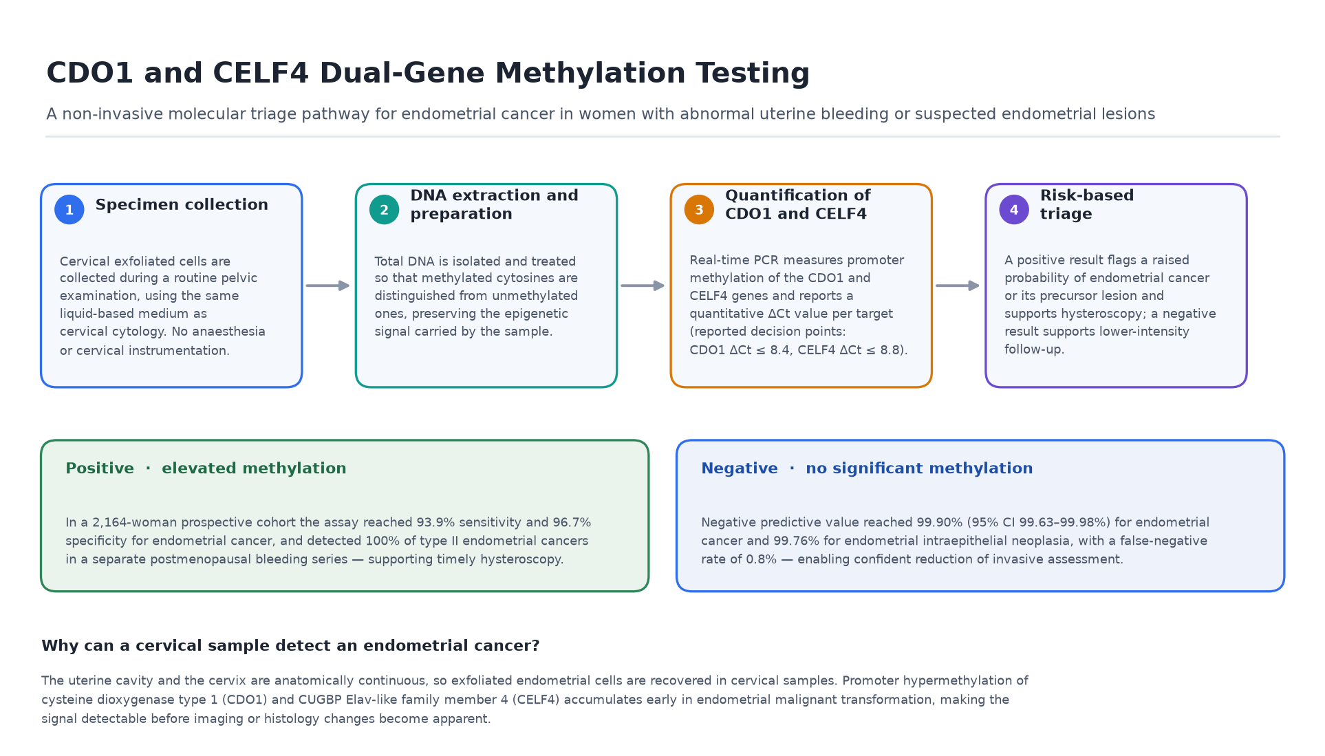
Summary — Endometrial cancer is the only gynaecological malignancy whose incidence is still rising worldwide, yet no non-invasive test is recommended for its detection in routine clinical practice; diagnosis still depends on hysteroscopy, dilatation and curettage, or endometrial biopsy. A DNA methylation assay that measures two gene targets — CDO1 (cysteine dioxygenase type 1) and CELF4 (CUGBP Elav-like family member 4) — in cervical exfoliated cells is changing that picture. Across ten independent cohorts published between 2017 and 2026, including a 2,164-woman prospective series and a 675-woman prospective study in Hong Kong, CDO1 and CELF4 methylation testing has delivered sensitivity of roughly 84%–94% and specificity of roughly 87%–99% for endometrial cancer, with negative predictive values approaching 99.9%. The test requires only the same cervical sample already collected for cytology, and it outperforms transvaginal ultrasound and serum CA125 in the same populations.
Key takeaways
- No new sampling procedure. The assay runs on cervical exfoliated cells collected with a routine liquid-based cytology sample — the uterine cavity and cervix are anatomically continuous, so exfoliated endometrial cells are recovered from the cervix.
- High specificity is the decisive advantage. Reported specificity reaches 96.7%–98.8%, against 45%–75% for transvaginal ultrasound and cytology combinations, which is what allows invasive assessment to be avoided.
- Very high negative predictive value. In the largest prospective cohort, NPV was 99.90% (95% CI 99.63–99.98%) for endometrial cancer and 99.76% for endometrial intraepithelial neoplasia.
- Works where ultrasound is weakest. 55.2% of premenopausal women with endometrial cancer had an endometrial thickness below the 11 mm threshold yet tested positive; 52.7% of postmenopausal women without cancer had a thickness at or above 5 mm yet tested negative.
- The strongest independent predictor. In multivariable analysis the methylation result carried an adjusted odds ratio for endometrial cancer of 103.9 in premenopausal and 118.0 in postmenopausal women (p < 0.001) — higher than any clinical or imaging variable in the model.
- Detects the hardest cancers. 100% of type II endometrial cancers in a postmenopausal bleeding cohort were detected, and methylation testing flagged three endometrial cancers and one cervical cancer that were only diagnosed histologically one to four months later.
The clinical gap, and why CDO1 and CELF4
Endometrial cancer is now the most common gynaecological malignancy in high-income countries. There is no internationally endorsed population-level screening programme, and the diagnostic pathway has remained stubbornly invasive: transvaginal sonography as the initial investigation, followed by hysteroscopy, endometrial biopsy, or dilatation and curettage. Both steps have drawbacks. Transvaginal ultrasound is inexpensive but its specificity is limited — reported between 24.3% and 74.0% — it performs poorly at the diagnostic threshold in premenopausal women, and sensitivity falls in patients with obesity, who are also the group at highest risk. Invasive sampling is inconvenient, costly, requires instrumentation and, in some patients, anaesthesia.
The clinical problem is one of triage. Abnormal uterine bleeding and postmenopausal bleeding are common and highly non-specific: after full evaluation, only about 0.33% of women presenting with abnormal uterine bleeding and approximately 9% of postmenopausal women are ultimately diagnosed with endometrial cancer. A test that reliably identified the small minority who need hysteroscopy, while confidently discharging the large majority, would remove a substantial volume of invasive procedures without delaying diagnosis.
DNA methylation supplies exactly that kind of signal. Aberrant promoter hypermethylation is an early and stable event in malignant transformation, detectable before histological or radiological changes are evident. Two markers have been repeatedly validated in endometrial carcinogenesis. CDO1 encodes cysteine dioxygenase type 1, an enzyme involved in cysteine metabolism and redox regulation whose promoter silencing is a well-documented early epigenetic event in endometrial and other tumours. CELF4 encodes a CUGBP Elav-like RNA-binding protein that regulates transcript stability and translation. Together they form a two-gene panel that has been repeatedly shown to discriminate malignant endometrial lesions from benign causes of bleeding. Notably, the published series converge on the same quantitative interpretation thresholds — CDO1 ΔCt ≤ 8.4 or CELF4 ΔCt ≤ 8.8 — which is unusual in molecular diagnostics and supports reproducibility across laboratories and cohorts.
Diagnostic performance: what the evidence shows
The evidence base is unusually broad for a novel molecular test, spanning single-centre and multicentre prospective cohorts, premenopausal and postmenopausal populations, and both bleeding-driven and lesion-driven referral pathways. Reported sensitivity for endometrial cancer ranges from 84.1% to 93.9% and specificity from 86.6% to 98.8%.
| Cohort / population | n | Sensitivity | Specificity | Source |
|---|---|---|---|---|
| Suspected endometrial lesions (prospective, hysteroscopy-confirmed) | 2,164 | 93.9% | 96.7% | Diagnostics 2026;16(2):174 |
| Reproductive-age women with abnormal uterine bleeding | 517 | 91.7% | 88.8% | Chin J Lab Med 2023;46(4) |
| Abnormal uterine bleeding | 216 | 91.2% | 96.7% | J Lanzhou Univ (Med Sci) 2024;50(7) |
| Postmenopausal women undergoing diagnostic hysteroscopy | 573 cohort | 91.7% | 91.8% | Gynecol Oncol 2026;206 |
| Premenopausal women undergoing diagnostic hysteroscopy | 573 cohort | 89.7% | 93.0% | Gynecol Oncol 2026;206 |
| Postmenopausal bleeding | 138 | 87.5% | 95.9% | CytoJournal 2024;21:15 |
| Postmenopausal women with suspected endometrial lesions | 143 | 87.5% | 90.8% | Natl Med J China 2023;103(12) |
| Premenopausal women with abnormal uterine bleeding | 296 | 85.7% | 87.6% | Int J Gynecol Cancer 2024 |
| Women with endometrial biopsy indications (cancer plus atypical hyperplasia) | 607 | 84.9% | 86.6% | Front Oncol 2023;13 |
| Prospective gynaecology clinic cohort (Hong Kong, China) | 675 | 84.1% | 98.8% | Cancers 2025;17(18):3010 |
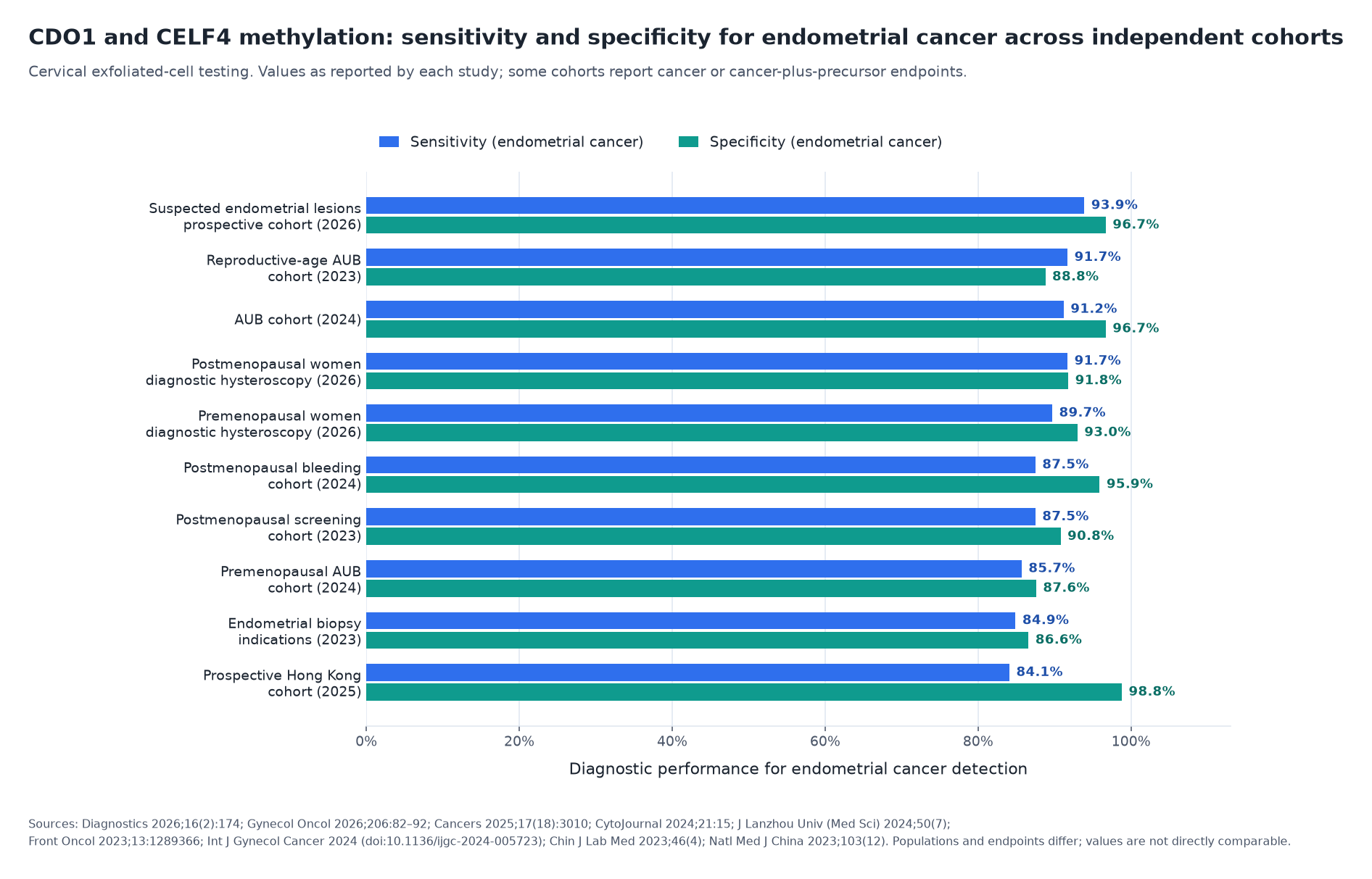
Three findings stand out. First, the largest prospective series — 2,164 women scheduled for hysteroscopy at a single tertiary centre, of whom 33 had endometrial cancer and 31 had endometrial intraepithelial neoplasia — reported 93.94% sensitivity and 96.7% specificity for cancer, with an NPV of 99.90% and an NPV of 99.76% for the precursor lesion. Second, the 675-woman Hong Kong prospective cohort, which compared methylation results against same-day endometrial histology, achieved an overall accuracy of 97.3%, specificity of 98.8%, PPV of 89.2%, AUC of 0.92 and a false-negative rate of only 0.8%. Third, in the 573-woman cohort stratified by menopausal status, adding endometrial thickness to the methylation result raised sensitivity to 96.6% in premenopausal and 100% in postmenopausal women — while methylation testing alone remained the dominant independent predictor in multivariable analysis.
Clinical scenarios and target populations
1. Women with abnormal uterine bleeding. This is the highest-volume clinical problem and the most valuable triage opportunity. In a 517-woman reproductive-age cohort, methylation testing achieved an AUC of 0.90 (95% CI 0.83–0.97) — the highest of any non-invasive modality assessed — with 91.7% sensitivity and 88.8% specificity, and outperformed age, body mass index, endometrial thickness and serum CA125. In a separate 216-woman cohort, sensitivity and specificity were 91.2% and 96.7%, against 55.9% and 78.6% for ultrasound and 32.4% and 76.4% for CA125.
2. Postmenopausal bleeding. Postmenopausal bleeding accounts for roughly two-thirds of gynaecological visits in postmenopausal women and affects up to 10% of them, yet only a minority prove to have cancer. In a 138-woman postmenopausal bleeding cohort, methylation testing achieved 87.5% sensitivity and 95.9% specificity, and detected 100% of type II endometrial cancers — the aggressive, non-oestrogen-dependent subtype that typically presents without endometrial thickening and carries the worst prognosis. In a separate 143-woman postmenopausal screening cohort, sensitivity was 87.5% and specificity 90.8%, rising to 100% sensitivity (95% CI 93.6–100%) when combined with transvaginal ultrasound.
3. Premenopausal women, where ultrasound is least reliable. Premenopausal endometrial assessment is confounded by cyclical thickness variation, and the diagnostic threshold is poorly specific. The 573-woman cohort found that 55.2% of premenopausal women with endometrial cancer had an endometrial thickness below 11 mm but tested methylation-positive, while 52.7% of postmenopausal women without cancer who had a thickness at or above 5 mm tested negative. In 296 premenopausal women with abnormal uterine bleeding, dual-gene methylation reached 85.7% sensitivity and 87.6% specificity, with an odds ratio of 42.53 (95% CI 11.90–152.04) — and combined with body mass index or endometrial thickness improved screening efficiency further.
4. Patients being referred for hysteroscopy. Used in the work-up of women already on a diagnostic pathway, the test can direct invasive assessment to those who need it. In a 607-woman cohort of patients with endometrial biopsy indications, methylation testing plus endometrial thickness achieved 94.9% specificity for endometrial cancer and atypical hyperplasia, against 86.6% for methylation alone. The Hong Kong cohort concluded explicitly that the assay “can act as a triage to reduce invasive endometrial assessment”.
5. Detection of precursor lesions. The assay is not limited to invasive cancer. Endometrial intraepithelial neoplasia was detected with 83.87% sensitivity, 97.95% specificity and an NPV of 99.76% in the 2,164-woman cohort — relevant because identifying and treating precursors is what prevents progression.
6. A signal that precedes histology. In the Hong Kong cohort, all seven false-positive cases proved to be neoplastic processes within the genital tract: five had endometrial hyperplasia and two had cervical intraepithelial neoplasia. Over the following one to four months, three of those patients were diagnosed with endometrial cancer and one with cervical cancer. The authors concluded that the assay “may be useful for detecting cancer early, before histological change is evident” — a property of genuine clinical rather than merely statistical interest.
Clinical value: a non-invasive alternative to an invasive default
The value proposition rests on a simple structural problem. Endometrial cancer has no recommended non-invasive screening test, so symptomatic women are investigated invasively. Yet only a fraction prove to have cancer. A test that is applied to a sample already being collected, that carries a negative predictive value above 99.7%, and that costs a fraction of hysteroscopy, can reduce procedure volume, waiting lists and patient discomfort without displacing histological confirmation where it is needed.
The comparative data support this. In the 517-woman cohort, methylation was better than every other non-invasive option; the same conclusion was reached in the 296-woman and 216-woman cohorts and in the 607-woman series. In the 573-woman cohort, the test was the single strongest independent predictor of endometrial cancer in both menopausal strata, with adjusted odds ratios of 103.9 and 118.0. And when combined with transvaginal ultrasound, sensitivity approached or reached 100% — a combination strategy that is practical precisely because ultrasound is already performed.
The foundation for the two-gene panel was laid by an earlier integrated epigenomics study, which screened methylomics databases and validated candidate markers in 146 cervical scrapings. That work identified CDO1 and CELF4, alongside one further gene, as the best-performing markers, with a two-of-three panel reaching 91.8% sensitivity, 95.5% specificity and an odds ratio of 236.3 (95% CI 56.4–989.6). That the same two targets have since been reproduced across independent cohorts in different provinces and in Hong Kong, China — with a common interpretation threshold — is what distinguishes a validated assay from a promising single-centre observation.
Appropriate use and honest limitations
Responsible communication requires stating clearly what this test is and is not.
It is a triage and adjunctive tool, not a replacement for histology. A positive methylation result indicates elevated probability of endometrial cancer or a precursor lesion and supports referral for hysteroscopy; it is not a diagnosis, and tissue confirmation remains the standard. A negative result substantially lowers risk but does not eliminate it.
The positive predictive value is modest in low-prevalence settings. In the 2,164-woman cohort, PPV was 31.0% — because endometrial cancer was uncommon among women referred with suspected lesions. This is the expected behaviour of any high-specificity test in a low-prevalence population, and it is why the test is best used to rule out rather than to rule in, and ideally in combination with clinical risk factors and imaging.
Cohorts differ and endpoints are not uniform. Some studies report endometrial cancer alone, others cancer plus atypical hyperplasia or endometrial intraepithelial neoplasia; sensitivity is therefore not directly comparable between rows of the table above. Several cohorts are single-centre and hospital-based, and the largest prospective series are drawn from Chinese and Hong Kong, China populations. Performance in other health systems, and in genuinely unselected populations rather than symptomatic referrals, requires further prospective evaluation.
Pre-analytical conditions matter. Methylation results were shown to be affected by endometrial thickness, abnormal cervical cytology and the presence of endometrial hyperplasia. Reported interpretation thresholds (CDO1 ΔCt ≤ 8.4, CELF4 ΔCt ≤ 8.8) assume standardised specimen handling, DNA extraction and real-time PCR conditions; laboratories adopting the assay must validate their own workflow before clinical reporting.
Frequently asked questions
What does the CDO1/CELF4 methylation test measure? It measures the level of DNA methylation at the promoter regions of the CDO1 and CELF4 genes in cervical exfoliated cells, reported as a quantitative ΔCt value for each target. Rising methylation indicates an increasing likelihood of endometrial malignant or pre-malignant change.
Why can a cervical sample detect a cancer of the uterus? The uterine cavity and the cervix form a continuous anatomical channel, so exfoliated endometrial cells are carried down and recovered in cervical samples. This is the same principle that allows cervical cytology to occasionally detect endometrial abnormalities.
Is a separate appointment or procedure needed? No. The sample is the cervical exfoliated cell specimen already collected in liquid-based medium during a routine gynaecological examination. No additional instrumentation or anaesthesia is required.
How does it compare with transvaginal ultrasound? Ultrasound is inexpensive and widely available but its specificity is limited, and its diagnostic threshold performs poorly in premenopausal women and in patients with obesity. Methylation testing has shown substantially higher specificity, and the two are complementary: combining them raised sensitivity to 96.6%–100% in the menopausal-stratified cohort.
Which women benefit most? Women with abnormal uterine bleeding or postmenopausal bleeding, premenopausal women in whom ultrasound is inconclusive, women at elevated risk through obesity, diabetes or unopposed oestrogen exposure, and any patient being considered for hysteroscopy in whom a non-invasive rule-out would be clinically useful.
Evidence base: peer-reviewed references
- [1] Yu Y, Su T, Zhang H, et al. Clinical validation of DNA methylation detection in cervical exfoliated cells for endometrial cancer in women with suspected lesions. Diagnostics. 2026;16(2):174. doi:10.3390/diagnostics16020174 (PMID 41594150).
- [2] Wang X, Zheng L, Zhu G, et al. CDO1 and CELF4 methylation assay as the dominant predictor of endometrial cancer: a cohort analysis across pre- and post-menopausal cohorts. Gynecol Oncol. 2026;206:82–92. doi:10.1016/j.ygyno.2026.01.776.
- [3] Lee HSJ, Wu S, Yeung SY, et al. Prospective evaluation of cervical scrapings CDO1 and CELF4 methylation assay in detection of endometrial cancer. Cancers. 2025;17(18):3010. doi:10.3390/cancers17183010.
- [4] Cai B, Du J, Wang Y, et al. The endometrial cancer detection using non-invasive hypermethylation of CDO1 and CELF4 genes in women with postmenopausal bleeding in Northwest China. CytoJournal. 2024;21:15. doi:10.25259/Cytojournal_78_2023.
- [5] Cai B, Li L, Liu B, et al. Clinical value of CDO1/CELF4 dual gene methylation detection in endometrial cancer screening for females with abnormal uterine bleeding. J Lanzhou Univ (Med Sci). 2024;50(7):28–36. doi:10.13885/j.issn.1000-2812.2024.07.004.
- [6] Qi B, Sun Y, Lv Y, et al. Hypermethylated CDO1 and CELF4 in cytological specimens as triage strategy biomarkers in endometrial malignant lesions. Front Oncol. 2023;13:1289366. doi:10.3389/fonc.2023.1289366.
- [7] Zhao X, Yang Y, Fu Y, et al. DNA methylation detection is a significant biomarker for screening endometrial cancer in premenopausal women with abnormal uterine bleeding. Int J Gynecol Cancer. 2024. doi:10.1136/ijgc-2024-005723 (PMID 39043572).
- [8] Zhao X, Xu D, Ma J, et al. Application of DNA methylation in detection of endometrial carcinoma in women with abnormal uterine bleeding at childbearing age. Chin J Lab Med. 2023;46(4):367–374. doi:10.3760/cma.j.cn114452-20221110-00670.
- [9] Kong L, Xiao X, Wan R, et al. The role of DNA methylation in the screening of endometrial cancer in postmenopausal women. Natl Med J China. 2023;103(12):907–912. doi:10.3760/cma.j.cn112137-20220929-02058 (PMID 36973218).
- [10] Huang RL, Su PH, Liao YP, et al. Integrated epigenomics analysis reveals a DNA methylation panel for endometrial cancer detection using cervical scrapings. Clin Cancer Res. 2017;23(1):263–272. doi:10.1158/1078-0432.CCR-16-0863 (PMID 27507616).
A note on this release
All quantitative statements above are drawn from the peer-reviewed publications listed in the reference section and are reported as published by their authors, including confidence intervals where available. Cohorts differ in menopausal status, presenting symptom, referral pathway and diagnostic endpoint — some report endometrial cancer alone, others cancer plus precursor lesions — so individual figures are not directly comparable across studies. This release describes the scientific and clinical evidence for CDO1 and CELF4 dual-gene methylation testing as a methodology; it deliberately contains no manufacturer or brand identifiers. Endometrial cancer assessment and any decision to perform hysteroscopy or endometrial biopsy should always follow local clinical guidelines and the judgement of the treating clinician.
Related reading: DNA methylation testing across oncology indications
This article is part of a series on the clinical use of DNA methylation testing. The other articles in the series, and the product page corresponding to the technology described in this article, are listed below.
- Cervical cancer: PAX1 and JAM3 dual-gene methylation testing for cervical cancer triage and the management of HPV-positive women
- Ovarian cancer: CDO1 and HOXA9 dual-gene methylation testing for ovarian cancer detection and pelvic mass triage
- Lung cancer: SHOX2 and RASSF1A DNA methylation PCR kit for the molecular triage of indeterminate lung lesions
- Colorectal cancer: Methylated SEPT9 DNA blood test for colorectal cancer screening and pre-colonoscopy triage
Related product: Human CDO1 and CELF4 Gene Methylation Detection Kit (Real-Time PCR)